Universität Regensburg
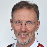
Reinhard Rachel

Anschrift:
Herr Prof. Dr. Reinhard Rachel
Universität Regensburg
Fakultät für Biologie und Vorklinische Medizin
Zentrum für Elektronenmikroskopie
Universität Regensburg
Fakultät für Biologie und Vorklinische Medizin
Zentrum für Elektronenmikroskopie
Straße:
Universitätsstr. 31
Ort:
93053 Regensburg
Tel.:
0941 943-2837
Fax:
0941 943-2868
Leistungsprofil:
Praxisrelevante Forschungsgebiete:- Extremophile Mikroorganismen (Archaeen, Bakterien, Viren)
- Ultrastruktur und Oberflächen von Nierengewebe, Zellen und Viren
- Transmissions-Elektronenmikroskopie, Raster-Elektronenmikroskopie; Raster-Transmission-Elektronenmikroskopie (STEM)
- 3D-Struktur-Analyse mit elektronenmikroskopischen Methoden; Immun-Elektronenmikroskopie
- Struktur von Membranen und Membranproteinen
Praxisrelevante aktuelle Projekte:
- Flagellen, Pili, Zell-Zell-Interaktion, Zell-Oberflächen-Interaktion bei extremophilen Mikroorganismen (Prof. Dr. Dina Grohmann, Dr. Robert Reichelt)
- Proteinkomplexe, Membranproteine und Oberflächenproteine von Archaeen und Bakterien (u.a. Prof. Dr. Christine Ziegler, Dr. Robert Reichelt)
- Ultrastruktur von Nierengewebe, Nierenzellen (Prof. Dr. Ralph Witzgall)
- Ultrastruktur von Mikroorganismen (Prof. Dr. Andreas Klingl, LMU München)
Praxisrelevante Ausstattung/Messmethoden:
- schonende Präparationsmethoden: Kryopräparation, Kryofixierung, Gefriersubstitution
- Gefrierbruch, Gefrierätzung, Schwermetall-Bedampfung: Cressington CFE-50; Kritisch-Punkt-Trocknung und Schwermetall-Besputtern
- Transmissions-Elektronenmikroskope (JEOL 200kV S/TEM, Tomographie-Einrichtung mit Doppelkippung, Kryohalter, sCMOS-Kamera); FEI/Philips CM12 mit Goniometer, Digital-Kamera)
- Ultramikrotomie
- digitale Bildverarbeitung, 3D-Rekonstruktion, Elektronentomographie
Publikationen:
- B.Daum, J.Vonck, A.Bellack, P.Chaudhury, R.Reichelt, SV Albers, R.Rachel, W.Kühlbrandt (2017) Structure and in-situ organization of the Pyrococcus furiosus archaellum machinery. eLIFE 6:e27470
- D Steppan, A Zügner, R. Rachel, and A Kurtz (2013) Structural analysis suggests that renin is released by compound exocytosis. Kidney International 83, 233-241
- R. Rachel, C. Meyer, A. Klingl, S. Gürster, T. Heimerl, N. Wasserburger, T. Burghardt, U. Küper, Bellack, S. Schopf, R. Wirth, H. Huber, G. Wanner (2010) Analysis of the ultrastructure of Archaea by electron microscopy. Methods in Cell Biology 96: 47-69
- U. Küper, C. Meyer, V. Müller, R. Rachel, H. Huber (2010) An energized outer membrane and spatial separation of metabolic processes in the hyperthermophilic Archaeon Ignicoccus hospitalis. Proc. Natl. Acad. Sci. USA 107: 3152-3156
- A. Veith, A. Klingl, B. Zolghadr, K. Lauber, R. Mentele, F. Lottspeich, R. Rachel, S.-V. Albers, and A. Kletzin (2009) Acidianus, Sulfolobus and Metallosphaera Surface Layers: Structure, Composition and Gene Expression. Molecular Microbiology 73: 58-72
- B. Junglas, A. Briegel, T. Burghardt, P. Walther, H. Huber, R. Rachel (2008) Ignicoccus hospitalis and Nanoarchaeum equitans: Ultrastructure, cell-cell interaction, and 3D reconstruction from serial sections of freeze-substituted cells and by electron cryotomography. Arch. Microbiol. 190, 395-408
- T. Burghardt, D.J. Näther, B. Junglas, H. Huber and R. Rachel (2007) The dominating outer membrane protein of the hyperthermopilic Archaeum Ignicoccus hospitalis: a novel pore-forming complex. Mol. Microbiol. 63: 166-176
- Ch. Moissl, R. Rachel, A. Briegel, H. Engelhardt, and R. Huber (2005). The unique structure of archaeal 'hami', highly complex cell appendages with nano-grappling hooks. Mol. Microbiol. 56: 361-370
- S. Nickell, R. Hegerl, W. Baumeister, and R. Rachel (2003) Pyrodictium cannulae enter the periplasmic space but do not enter the cytoplasm, as revealed by cryo-electron tomography. J. Struct. Biol. 141(1): 34-42
- R. Rachel, M. Bettstetter, B.P. Hedlund, M. Häring, A. Kessler, K.O. Stetter, and D. Prangishvili (2002) Remarkable morphological diversity of viruses and virus-like particles in hot terrestrial environments. Arch. Virology 147: 2419-2429
Kooperationsangebot für die Wirtschaft / Praxis:
Bevorzugte Form der Kooperation:
- Beratung
- Gutachten
- Messung
- FuE
- Bildung
Bestehende Kooperationen:
Mit Hochschulen:
Universität Ulm
Präp.-Methoden
seit 2000
Universität München
TEM, SEM, Präp.Methoden
seit 1998
University of Exeter
electron cryo-tomography
seit 2016
Mit Unternehmen:
Leica
Kryopräparation
seit 2006
JEOL
STEM Tomography
seit 2014
Sonstiges:
seit April 2022 im Ruhestand
Zurück zur Liste
Falls dies ihr Profil ist, können Sie es hier nach dem Login bearbeiten.
